For Non-Scientists
Welcome to the Fleischer Lab! This page is targeted to visitors with expertise outside of science and contains non-technical descriptions of our research. We invite you to explore our website and learn more about our work. Thank you for your interest and we welcome your questions and inquiries. Contact us HERE.

Who We are
We are a basic and translational research laboratory at the Emory University School of Medicine. Emory University is a private, not-for-profit institution located in the Historic Druid Hills neighborhood of Atlanta, Georgia. Emory University is a research intensive university according to the Carnegie Classification of Institutions of Higher Education (read more HERE). Basic research is focused on gaining a molecular-level understanding of how the world works. For our lab, that includes understanding the physics and engineering behind magnetic resonance imaging (MRI) scanners and determining how our brain is affected by disease. Translational research takes our findings from basic research and attempts to 'translate' them into clinics and hospitals.

what we research
Most of our research is focused on imaging and spectroscopy using magnetic resonance imaging (MRI) scanners. MRI uses radiofrequency waves (which are not harmful) to generate images of the body and brain. We also use magnetic resonance spectroscopy (MRS), a similar technique that uses electromagnetic waves to provide chemical and molecular information. One of the earliest applications of spectroscopy is in space research. A nice description of spectroscopy used at NASA can be found HERE. Our research is a combination of developing new techniques and applying them to improve our understanding of the healthy and diseased brain. Further descriptions of three research projects are included below.
Cancerous Brain Tumors
The Centers for Disease Control and Prevention estimate over 22,000 new brain cancer diagnoses are made in the U.S. each year. Gliomas are a type of brain cancer that account for nearly 75% of all cancerous brain tumors. The most aggressive form, called glioblastoma, has a five-year survival rate of less than 6% (Central Brain Tumor Registry of the US). One of the hallmarks of all cancers is an increase in inflammatory markers – small molecules released by the body in response to illness or injury. We observed that systemic inflammation can lead to changes in brain chemistry, particularly increases in glutamate (Read more HERE). Our goal is to identify new inflammation-related biomarkers, or indicators, that can be used to monitor brain tumors and their treatment. We are developing new non-invasive tools for MRI scanners to monitor tumor growth and response to treatment, with the end goal of increasing survival rates for brain tumor patients.


Brain Thermometers
Brain temperature is important for healthy brain activity and recovery after brain injury. Currently, the only clinical method that exists to measure brain temperature is an invasive, surgically-implanted probe inserted into the brain through the skull and is impractical for routine use in healthy humans. We are developing new, non-invasive methods for measuring brain temperature using MRI scanners. These tools are being tested in both healthy human volunteers and in patients after head injury or disease such as stroke or concussion.
New Technology for MRI scanners
MRI scanners are commonly used in hospitals and outpatient clinics. The benefits of MRI and a similar technique, MRS, are that these methods are non-invasive (when used without contrast agents), provide high-resolution images of the brain and body, and can be repeated to look at changes over time. However, there is still room for improvement. Many MRI techniques are slow and patients have to lie in the scanner for long periods of time. Motion often distorts MRI images and can be difficult to correct. We are working to develop new methods for acquiring and analyzing data from MRI scanners.
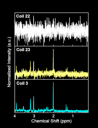
MR spectra from 3 different radiofrequency coils in a multi-channel head array for acquiring brain data.

